

This term ‘RCT’ has instilled more fear in people than any other dental procedure. We very often get to hear that it is a dreadful experience, it is very very painful, they dig and drill the tooth, Pain lasts for over many weeks to days, so on and so forth.
Well, let us help you understand this RCT or Root canal therapy.
What is a ‘RCT’ ? To answer this we must first take a look at your tooth itself.
The human teeth is made up of ENAMEL, DENTINE, and PULP. The outer hard and rigid structure is called ENAMEL that surrounds “DENTINE ” which is also hard in nature and makes up the bulk of tooth structure. Dentine consists of small fluid filled tubules (Dentinal tubules) that extends from the inner most pulp chamber (that houses the pulp tissue) to the enamel. PULP is present in the central portion of the tooth and consist of connective tissue with cells blood vessels ,nerves , lymphatics etc.
PULP is many ways is like the heart of the tooth. It is protected from the microbial rich oral environment by Enamel and Dentine. Pulp can be divided in to 2 parts.
When tooth decay progresses unchecked and breaches the enamel and dentin barrier , it leads to inflammation of the dental pulp. This is an extremely painful experience and requires immediate dental treatment ( Emergency dental care).
The only way to treat a tooth in such a situation is to remove the dental pulp with all its blood vessels and nerve connections. The procedure of removing both the coronal pulp and radicular pulp is called Root canal therapy.
ROOT CANAL THERAPY or RCT , comes under the purview of Endodontics. And typically a specialist who is an endodontist or a dentist trained adequately to perform RCT , can do RCT!
Now that we have established what is RCT , lets move ahead and answer what is done during the procedure. lets break it down for you.
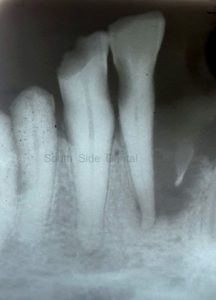
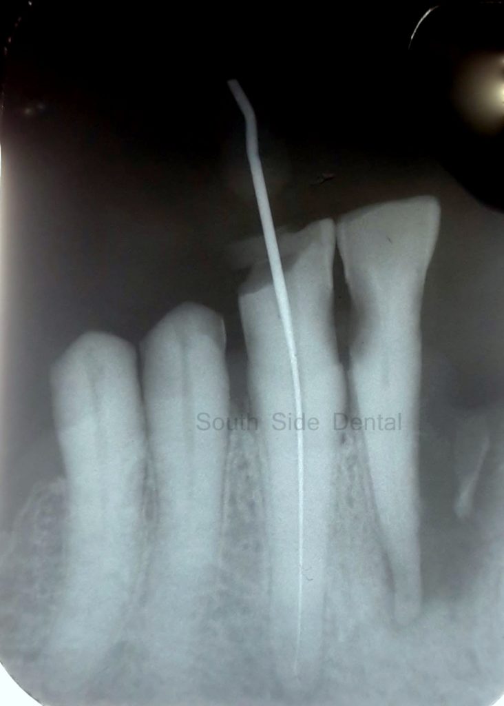
Step 1: X ray showing pulpally infected lower left canine
STEP 1. Before doing the procedure. We take our time to do a few tests to ascertain that the tooth really does need a RCT. If not a simple filling will solve the problem. Diagnostic X rays are taken to see how far deep into the tooth the decay has progressed and how close is it to the dental pulp. Many times additional test to check the vitality of the pulp many be needed. We may use a cold test or a heat test to confirm that the tooth is pulpaly infected or many time non vital ( Dead).
STEP 2. Local anesthesia. This the part where we ensure that the tooth under consideration is completely numb. Profound local anesthesia is essential to successful outcome. We need you to be comfortable on the chair. This is where things become difficult for many patients who have previously had RCT done and they recollect their experience and painful episodes during the RCT appointments.
The cause for all fear during the RCT.
If anesthesia has not worked in your case then the procedure can become painful. Before making any access in to your tooth we make sure that the tooth is numb . If not more anesthesia is provided.
Occasionally an infected tooth, that has collected a lot of pus (Perapical Abscesss) may not respond well to local anesthesia given through infiltration technique. Such a situation needs Nerve block technique of anesthesia or more anesthesia. Pre-medication with a pain killer can help relieve anxiety during the procedure.
A rubber dam or an tooth isolating sheet is then placed on top of the tooth to be treated, This isolates the tooth from the oral cavity and prevent contamination with saliva. It also protects the oral cavity from chemicals that are used during the procedure.
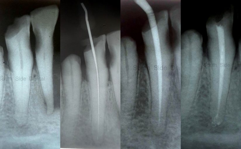
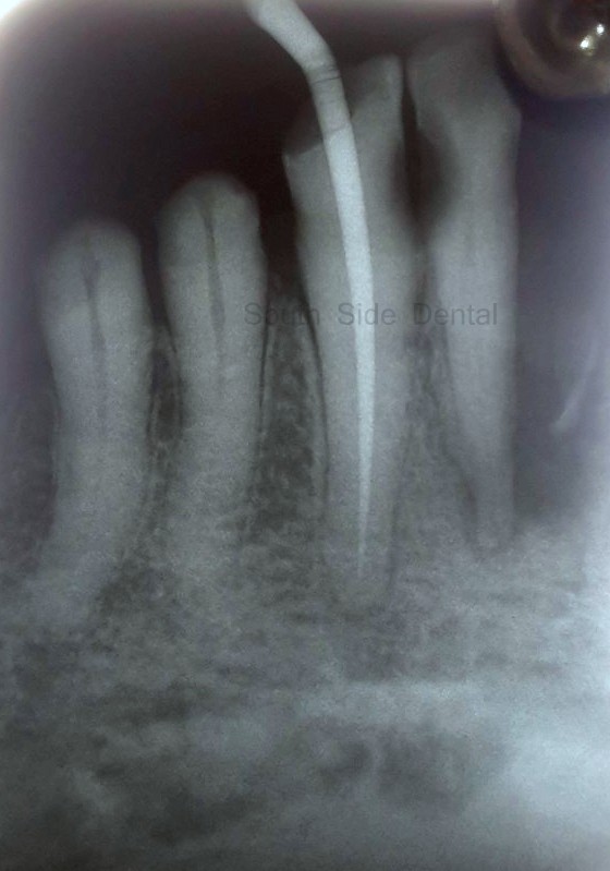
Step 3: Endodontic access
STEP 3. We make an access in to the tooth to reach in to the pulp chamber. The access is then prepared to enable us to use endodontic or RCT specific instruments. These instruments are highly flexible and can move deeper in to the tooth tooth providing access in to the root canal.
STEP 4. Once access is gained the next step is to completely clean and shape the canal of the tooth using the instruments and chemicals approved for this procedure. The canal must be free of all remnants of the infected pulp. The most potent of these chemical is the Sodium Hypochorite. It is also very caustic and can cause chemical injury if used carelessly with out the protection of the rubber dam.
STEP 5. If the tooth is highly infected then the we may require more than one appointment to complete the root canal. Long standing infected tooth (Chronic) need more than one appointment. If such is the case then a medicament is placed inside the prepared canals and temporarily sealed for a week or two.
Many a times the following step can effectively be completed in the same appointment ( Single sitting RCT)
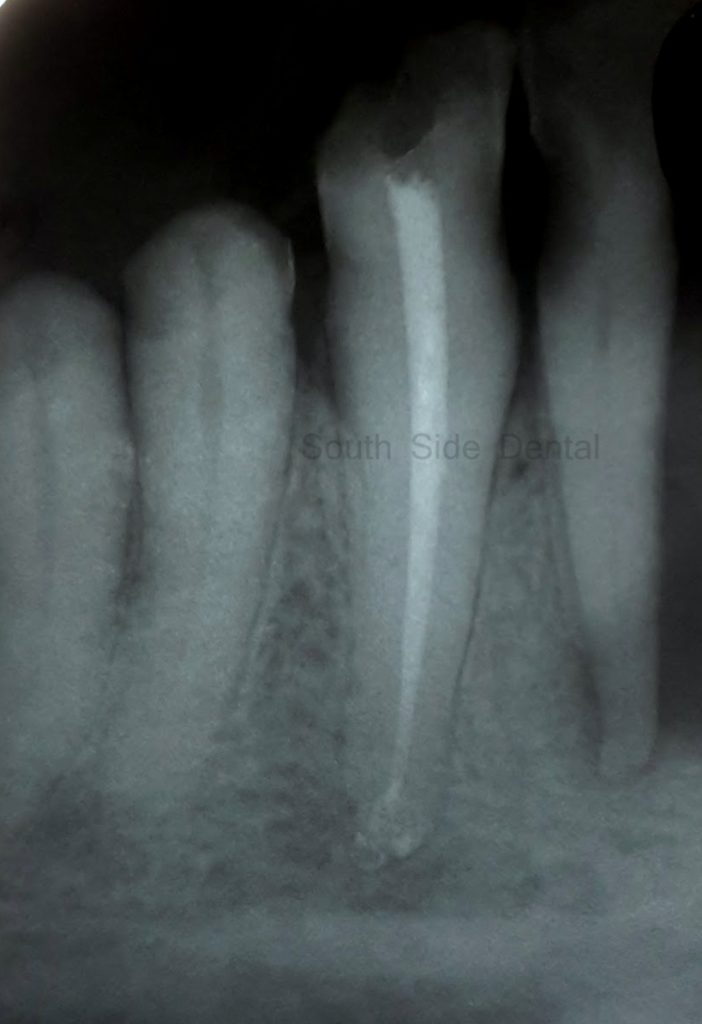
Step 6: Obturation or root canal filling
STEP 6. The final step : Obturation of sealing of the prepared disinfected root canals. The material of choice to fill the canals is actually made froma bark of a tree. It is called ‘Gutta Percha'(GP). Reinforced with other materials to provide strength, the GP is placed in the canal and sealed using a special root canal sealer material.The GP is an inert material and perfect for root canal filling.
STEP 7. Once the root canal is filled , it is time to fill the pulp chamber. Sometimes the filling of the pulp chamber is done a day or two after obturation.
The filling of the pulp chamber is referred to as Core Filling. Composite or tooth colour material is often used these day. Many prefer to use Silver Amalgam as a filling. Southside dental is an Amalgam / mercury free clinic. We prefer to use composites as core filling or tooth filling materials.
Step 6: Final obturation picture
STEP 8. It is not always necessary to provide a full coverage cap or crown for root canal treated teeth. However if the decay has involved multiple surfaces compromising the tooth support, then it is advisable to provide a cap or crown for the tooth.
The entire process from step 1 to 8 may take any where between 2 to 4 weeks. It can be done sooner too is some situations. However we believe that BIOLOGY must not be rushed.
Hope this blog gives you a better understanding of what the allegedly dreaded RCt is all about and what goes in to doing this specialty procedure. Hope you come over the fear of RCT and get timely help.
Prevent dental problems by visiting your dentist regularly.
FEAR THE DISEASE not THE DENTIST

Dr.Jayanthi Kumaresh
Teeth is something that makes our smile special. Oour dentist is someone who makes us smile with grace and confidence.
Read More
Sagar Kanta
Toothache can actually cause depression.. if you don’t happen to meet a good dentist. But if you have a great dentist, then tooth problems can be as simple as common cold.
Read More
Sohail Chandan
An Amazing pain free experience by a very professional and capable team. They good care of me and always checked on me to see if I was facing any issues.
Read More
Subham Singh
The doctor is very friendly…! The way I was treated in the clinic was something unexpected from a dentist. I would recommend visiting him whenever you face any problem.
Read More
Bhuvana Ravikumar
Professionalism, Hygienic & Compassionate makes Dr.Niranjan & Dr.Deepti the best dentist doctor couple.
Read More